

- The majority of the cortex has 6 cortical layers
- The six layers from superficial to deep:
- Molecular
- External granular
- External pyramidal
- Internal granular
- Internal pyramidal (ganglion)
- Multiform.
- The six layers from superficial to deep:
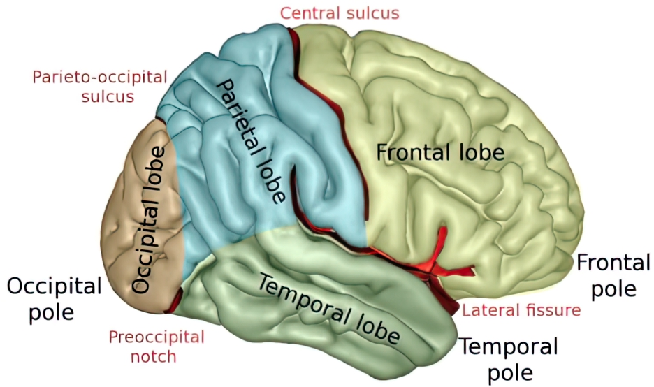
- The cortex is organized into 5 lobes: frontal, parietal, temporal, occipital, and limbic.
- Frontal lobe: Bound by the central sulcus and lateral fissure.
- Parietal lobe: Bound by the central sulcus, lateral fissure, and parieto-occipital fissure.
- Temporal lobe: Bound by the Sylvian fissure and preoccipital notch.
- Occipital lobe: Bound by the parieto-occipital sulcus and preoccipital notch.
- Limbic lobe: Consists of the parahippocampal, cingulate, and subcallosal gyri. The limbic lobe interacts with various other structures that are part of the limbic system.

Lobes of the brain
- The majority of the cortex has 6 cortical layers
Frontal Lobe
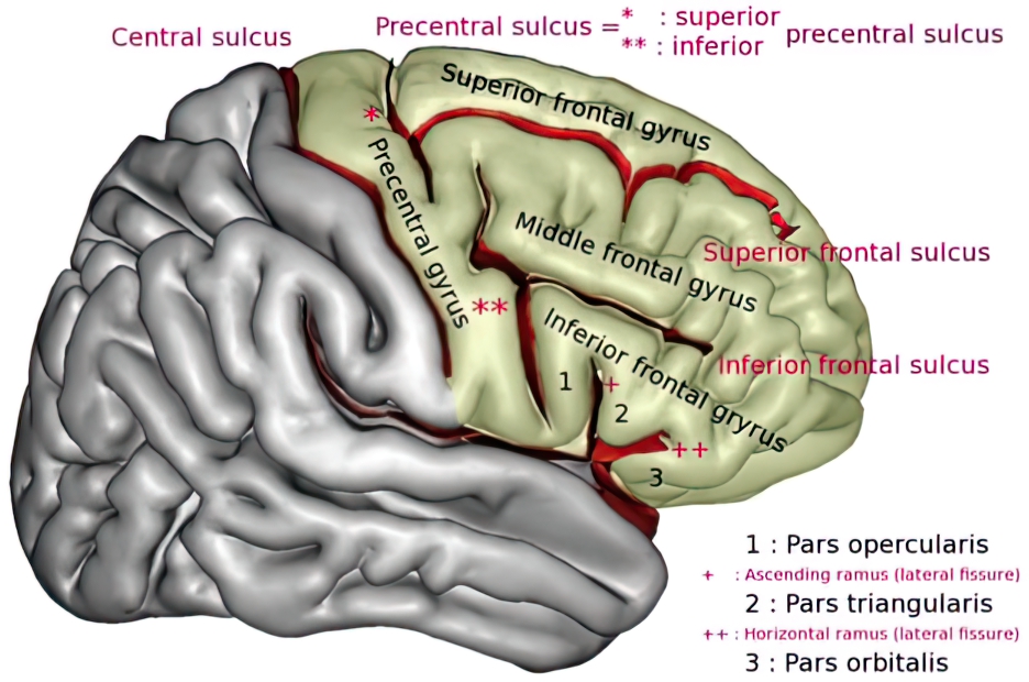
- There are four major gyri: precentral, superior frontal, middle frontal, and inferior frontal.
- The precentral gyrus is the primary motor strip.
- The motor homunculus of the precentral gyrus:
- The superior frontal contains the supplementary motor area (SMA).
- The middle frontal contains the frontal eye fields which are necessary for voluntary saccadic eye movements.
- The inferior frontal is organized into the pars orbitalis, pars triangularis, and pars opercularis.
- Pars opercularis and triangularis are associated with Broca’s area.
- Pars orbitalis has a role in thought, cognition, and planning behavior.
Parietal Lobe
- There are five principal parts: post-central gyrus, superior parietal lobule, inferior parietal lobule, the precuneus, and the posterior portion of the paracentral lobule.
- The postcentral gyrus contains the primary sensory cortex.
- The sensory homunculus of the postcentral gyrus:

- The superior parietal lobule contains the somatosensory association area.
- The inferior parietal lobule has two components: angular and supramarginal gyri
- This is the sensory association cortex and has a role in perception, vision, reading, and speech.
- A lesion to this region can lead to Gerstmann’s syndrome.
- The precuneus is an area of cortex just anterior to the occipital lobe on the medial surface. It has a broad spectrum of functions including visuospatial processing, memory, and first-person perspective.
- It is an early region of atrophy in Alzheimer’s dementia
- The posterior portion of the paracentral lobule is regarded as a tertiary somatosensory cortex involved in stereognosis.
- Stereognosis: the perception, understanding, recognition, and identification of an object by touch.
- Tested by having the patient feel an object and identify it, such as a paperclip or set of keys.
- It is often accompanied by other deficits like agraphesthesia.
- Stereognosis: the perception, understanding, recognition, and identification of an object by touch.
- Responsible for emotion, behavior, and long-term memory formation.
- The limbic system is comprised of multiple structures including the limbic lobe (parahippocampal, cingulate, and subcallosal gyri), amygdala, hippocampus, mammillary bodies, and anterior thalamus.
Papez Circuit
- The Papez circuit is a collection of structures of the limbic system that connect the limbic lobe and the hypothalamus.
Cingulate gyrus
- Lies immediately above the corpus callosum.
- Receives input from the thalamus and surrounding cortex.
- Projects fibers via the cingulum to the parahippocampal gyrus.
- Unilateral damage to the cingulate gyrus can lead to apathy or cognitive dysfunction. Bilateral damage can cause akinesis and mutism.
Parahippocampal gyrus
- Represents the cortex that surrounds the hippocampus.
- Axons from the parahippocampal gyrus project to the hippocampus.
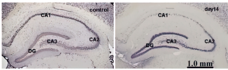
Hippocampus
- Located in the inferomedial temporal lobe.
- Associated syndromes:
- Gliosis and neuronal loss of the hippocampal CA1 pyramidal cell layer can lead to mesial temporal sclerosis (MTS) and focal epilepsy.
- Presents with focal seizures with impaired awareness.
- Area CA1 is particularly sensitive to ischemic damage
- Lesions to bilateral hippocampi can lead to profound anterograde amnesia.
- Gliosis and neuronal loss of the hippocampal CA1 pyramidal cell layer can lead to mesial temporal sclerosis (MTS) and focal epilepsy.
Fornix
- The major outflow white fiber tract from the hippocampus synapse at the mammillary bodies.
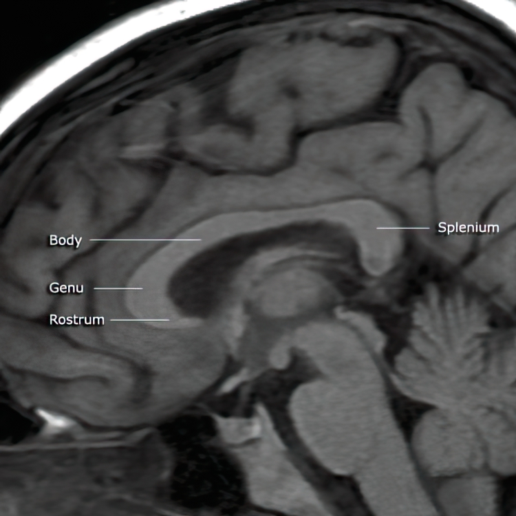
- Located below the splenium of the corpus callosum.
Mammillary bodies
- The mammillary bodies are round, paired structures located on the inferior surface of the hypothalamus.
- The mammillothalamic tract connects the mammillary nucleus to the anterior nucleus of the thalamus.
- Mammillary body dysfunction occurs in the setting of Wernicke’s encephalopathy.
Anterior nucleus of the thalamus
- The anterior thalamic nucleus projects to the cingulate cortex through the thalamocingulate fibers, completing the Papez’s circuit.
Temporal Lobe
- The lateral surface of the temporal lobe has three gyri: superior, middle, and inferior. They are separated by the superior, middle, and inferior sulci.
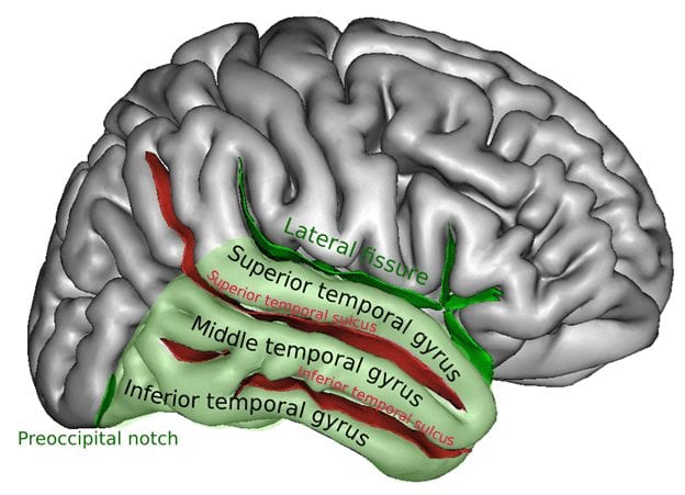
- The superior temporal gyrus is the region associated with language comprehension.
- When injured in the dominant hemisphere it leads to a Wernicke’s aphasia.
- The middle and inferior temporal gyri have a role in formed vision and processing.
- The fusiform gyrus (also called the occipitotemporal gyrus) is necessary for facial recognition.
- Lesions here can cause prosopagnosia/visual agnosia and Capgras delusion.
- Prosopagnosia: The failure to visually identify objects and faces.
- Capgras delusion is a variant of prosopagnosia and psychosis where the patient has the belief that close friends or family are replaced by an imposter.
- Lesions here can cause prosopagnosia/visual agnosia and Capgras delusion.
- There is an incredible number of connections organized throughout the brain, with several distinct axonal bundles that have well-defined functions.
U-fibers:
- Also called arcuate fibers, these link one gyrus to another.
- Most leukodystrophies (errors in the initial creation of myelin) will characteristically “spare the U-fibers” as opposed to a demyelinating process (damage to existing myelin) that will not.
Long association fibers
- Connect to different ipsilateral regions in the brain:
Arcuate fasciculus
- Links Wernicke’s and Broca’s areas.
- If damaged patients will develop conduction aphasia.
Commissural fibers
- Connect contralateral cerebral hemispheres:
Corpus callosum
- Organized into four segments; the rostrum, genu, body, and splenium.

Sagittal view of the corpus callosum on MRI - Associated syndromes:
- Alexia without agraphia: Occurs secondary to infarction of the splenium of the corpus callosum and left occipital lobe.
- Agenesis of the corpus callosum: A midline patterning defect that can be seen with other developmental abnormalities.
Anterior commissure
- Connects the olfactory bulbs, amygdala, and basal forebrain.
Posterior commissure
- Connects language processing centers from both hemispheres.
Hippocampal commissure
- Aides in memory formation.
Projection fibers
- Link the brain and spinal cord.
- They can be either afferent or efferent.
- The internal capsule is the most important.
- It is divided into three regions:
- Anterior limb: Has multiple different radiations but includes many thalamocortical fibers.
- Genu: Contains the corticobulbar tract (cortex to the brainstem).
- Posterior limb: Contains corticospinal (axons from the primary motor cortex), somatosensory, and corticopontine fibers.
- It is divided into three regions:
Log in to View the Remaining 60-90% of Page Content!
Important: If you signed up after 1/1/2026, or if you opted to migrate your old account to the new & improved platform (same great content, better experience), please log in at nowyouknowmed.com
