EMG and NCS are some of the mainstays of neurology diagnostic procedures. Interpretation and clinical correlation are imperative in order to be a great diagnostician and neurologist. Too, these skills will help you excel on the RITE, board exam, and in your career. For the medical student Shelf exam, it is not necessary to be able to interpret EMG/NCS in detail. In this chapter, you will explore EMG and NCS basic physiology and interpretation, and then test your retention at the end with our question bank! A handful of real EMG recordings have also been incorporated into the chapter. For more, check out our EMG Video Gallery and our EMG/NCS Case Bank.
Author: Brian Hanrahan MD
Chapter Multimedia Content
Introduction
Nerve conduction studies (NCS) and needle electromyography (EMG) are integral tools for the clinical neurologist and are considered to be an extension of the neurological examination. They can provide invaluable information to allow one to diagnose various peripheral nervous system (PNS) or muscle disorders. They can help localize a lesion (peripheral nerve, neuromuscular junction, muscle), discern the extent of the pathology (focal, multifocal, diffuse), and evaluate whether nerve-related pathology is axonal or demyelinating.
Basics
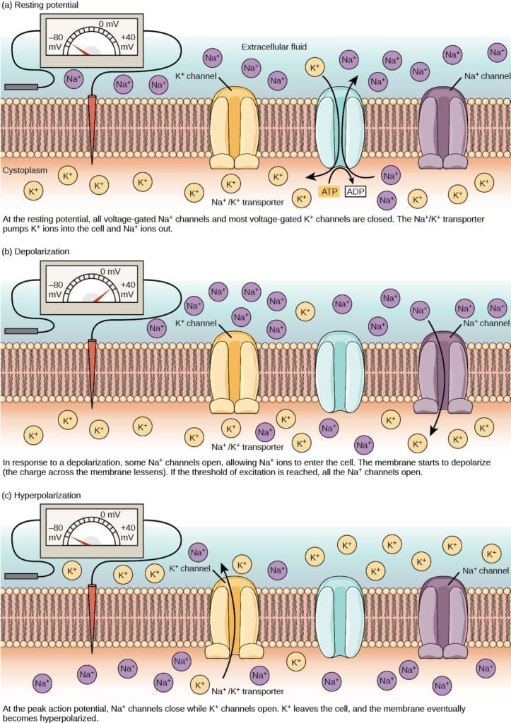
- Neurons have an intracellular resting membrane potential of -70 mV. When nerves conduct electrical impulses along their axon, the membrane is depolarized by an influx of sodium (Na+) ions via voltage-gated sodium channels (Figure 1).

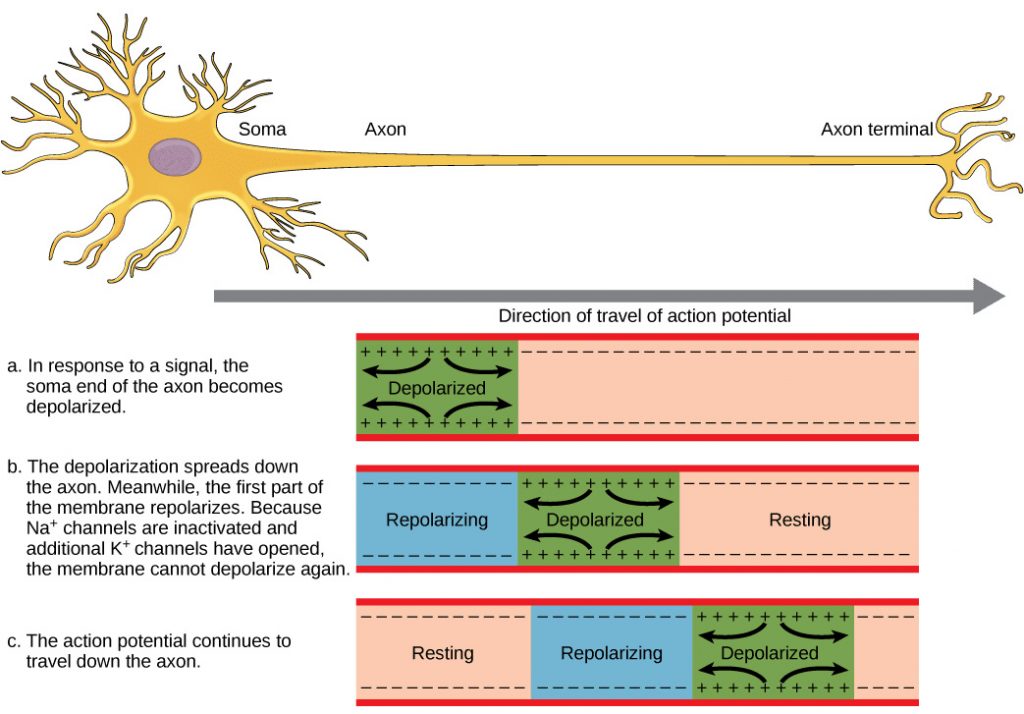
- This influx causes a voltage change to adjacent regions and changes the resting potential further down the axon thereby propagating the potential down the axon (Figure 2). The axon membrane potential will then stabilize with the inactivation of sodium channels and delayed opening of voltage-gated potassium channels.
Figure 2: A traversing action potential is conducted down the axon as the axon membrane depolarizes, then repolarizes.

- When unmyelinated, fibers can conduct potentials in the range of 1-5 m/sec. However, myelinated motor nerve axons and many sensory nerve axons have conduction velocities up to 150 m/sec. This is due to a process called saltatory conduction.
- Produced by Schwann cells, myelin sheaths are concentrically wrapped around an axon.
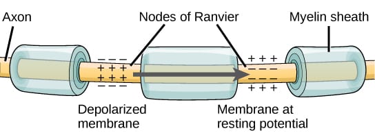
- In between myelin sheaths, there are gaps of exposed axon called nodes of Ranvier. It is at these nodes where action potentials occur.
- When an action potential occurs at a node, that current flows passively within the myelinated internode of the axon until the next node is reached.
- Saltatory Conduction: Current flows across the neuronal membrane and jumps from node to node (Figure 3).
Figure 3: Myelinated axon showing saltatory conduction

Log in to View the Remaining 60-90% of Page Content!
New here? Get started!
(Or, click here to learn about our institution/group pricing)1 Month Plan
Full Access Subscription-
Access to full question bank
-
Access to all flashcards
-
Access to all chapters & site content
3 Month Plan
Full Access Subscription-
Access to full question bank
-
Access to all flashcards
-
Access to all chapters & site content
1 Year Plan
Full Access Subscription-
Access to full question bank
-
Access to all flashcards
-
Access to all chapters & site content

