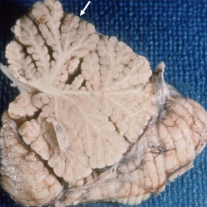
Below you will find over 200 high-yield neuropathology slides and images. These images were selected to be the most valuable for in-service/RITE* and ABPN board examinations.
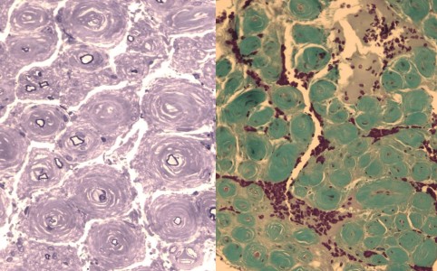
CIDP with Onion Bulbs
Medium power microscope slides; toluidine blue (left) and trichrome (right) stains of a peripheral nerve showing very large onion bulbs surrounding small thinly-myelinated nerve fibers secondary to repeated cycles of demyelination and re-myelination. This can be appreciated in chronic demyelinating neuropathies such as CIDP.
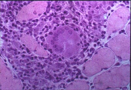
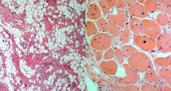
Sarcoidosis of Muscle
A skeletal muscle biopsy H&E stain with a non-caseating granuloma and a multinucleated giant cell.
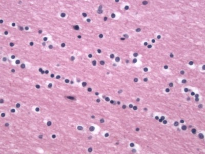
Normal Oligodendrocyte
Oligodendrocyte nuclei can be seen in white matter, often in short chains, and have smaller more dense nuclei than astrocytes.
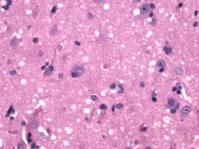
Spongiform Encephalopathy (CJD)
H&E stain showing many small round vacuoles consistent with spongiform encephalopathy.
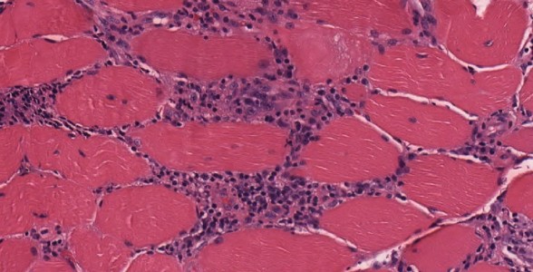
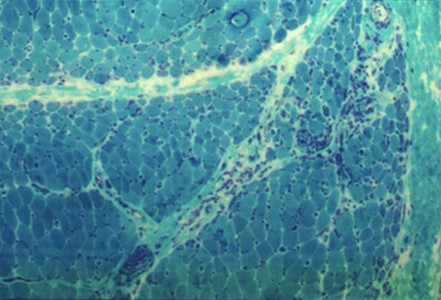
Polymyositis
Muscle biopsy with inflammatory cells in the endomysium (between and around individual myofibers) consistent with polymyositis.
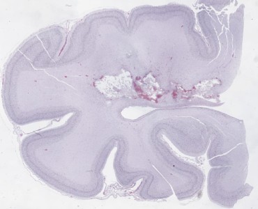
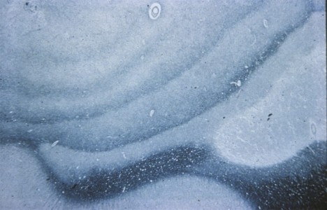
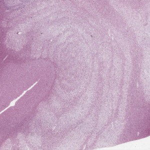
Periventricular Leukomalacia
Very low power view poorly-stained H&E with chalky white periventricular white matter lesions with necrosis.
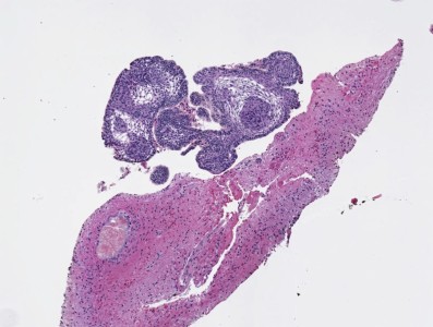
Craniopharyngioma
Encapsulated tumor with well-differentiated non-keratinizing squamous epithelium and papillary fibrovascular stroma.
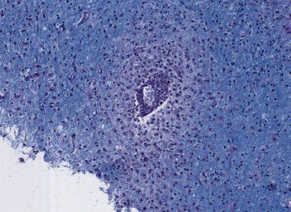
Acute Disseminated Encephalomyelitis
Biopsy with perivenous infiltration of lymphocytes and demyelination.
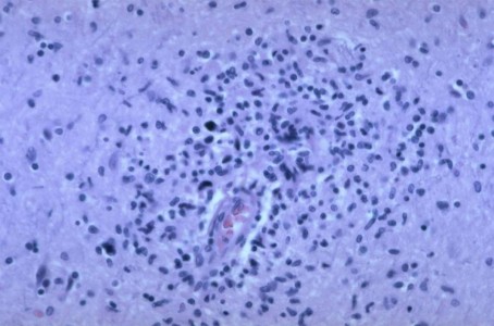
HIV Brain Biopsy
Brian biopsy with perivascular macrophages and multinucleate giant cells in white matter.
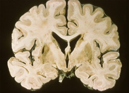
Lacunar Stroke in the Putamen
Note the small lake-like vacuous space within the putamen on the left side of this image.
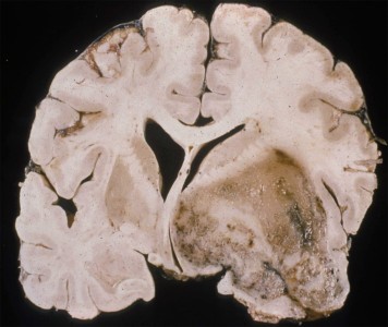
Glioblastoma Multiforme
Coronal gross cut. Large necrotic mass with increased vascularity and mass effect on the surrounding white matter.
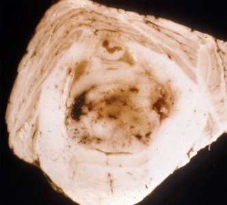
Medulloblastoma
Cerebellar mass with mass effect into the 4th ventricle consistent with a medulloblastoma.
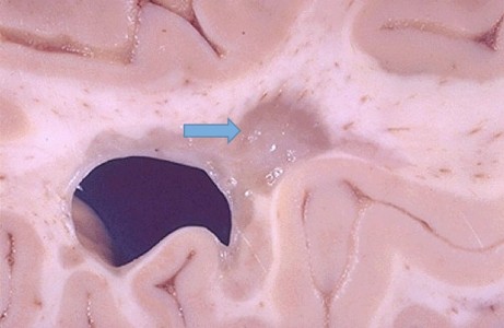
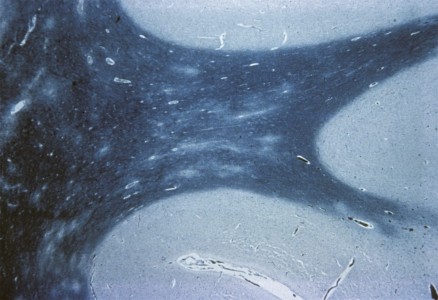
Multiple Sclerosis Plaque
Note the well-demarcated periventricular tan-gray patches of demyelination (arrow).
Log in to View the Remaining 60-90% of Page Content!
New here? Get started!
(Or, click here to learn about our institution/group pricing)1 Month Plan
Full Access Subscription
$142.49
$
94
99
1 Month -
Access to full question bank
-
Access to all flashcards
-
Access to all chapters & site content
3 Month Plan
Full Access Subscription
$224.98
$
144
97
3 Months -
Access to full question bank
-
Access to all flashcards
-
Access to all chapters & site content
1 Year Plan
Full Access Subscription
$538.47
$
338
98
1 Year -
Access to full question bank
-
Access to all flashcards
-
Access to all chapters & site content
Popular


